Tucked away in the heart of Philadelphia sits a museum unlike any other, where human bones, preserved organs, and medical oddities fill every glass case. The Mütter Museum isn’t your typical afternoon outing, but it offers a fascinating glimpse into medical history that you won’t find anywhere else.
From towering skeletal displays to jars containing mysterious specimens, this collection challenges visitors to confront the strange and sometimes unsettling realities of the human body. Ready to discover what makes this place so unforgettable?
1. The Tallest Skeleton In North America
Standing at an incredible seven feet six inches tall, the skeleton of a man who suffered from gigantism towers over museum visitors like a gentle giant frozen in time. His bones tell a story of struggle, as the condition caused by excess growth hormone created both physical challenges and social isolation during his lifetime. Doctors believe he lived during the 1800s, when medical science couldn’t offer the treatments available today.
Walking up to this display feels surreal because you truly grasp how massive this person was compared to average humans. The vertebrae, leg bones, and skull are all proportionally enlarged, showing how the entire body responded to hormonal imbalance. Museum guides explain that people with gigantism often experienced joint pain and heart problems.
Visitors often pause here the longest, snapping photos and contemplating what daily life must have been like for someone of such extraordinary height. Historical records suggest he may have worked in exhibitions, displaying himself to curious crowds. This skeleton remains one of the museum’s most photographed and discussed specimens.
2. The Soap Lady’s Preserved Body
One of the museum’s most famous and eerie exhibits features a woman whose body underwent a rare chemical transformation after burial. Her remains converted into a waxy, soap-like substance called adipocere through a process that occurs when body fat reacts with water and alkaline soil. Scientists estimate she died sometime in the early 1800s in Philadelphia.
The preservation is so complete that facial features, hair, and body shape remain visible after nearly two centuries. Museum experts explain that this natural mummification happens only under specific environmental conditions, making the Soap Lady an exceptionally rare specimen. Her yellowish-white appearance gives her an otherworldly quality that both fascinates and unsettles visitors.
Researchers have studied her remains extensively to learn about burial practices, body decomposition, and historical disease patterns. Some visitors find the display respectful and educational, while others debate the ethics of exhibiting human remains. Regardless of personal feelings, the Soap Lady represents an incredible example of natural preservation that continues teaching medical students and curious minds alike about post-mortem chemistry.
3. The Wall Of Skulls With Medical Conditions
Imagine walking into a room where hundreds of human skulls line the walls from floor to ceiling, each one telling a different medical story. This massive collection, known as the Hyrtl Skull Collection, contains 139 skulls gathered by Viennese anatomist Joseph Hyrtl in the 1800s. He collected them to disprove the racist pseudoscience of phrenology, which falsely claimed skull shape determined intelligence and character.
Each skull displays unique characteristics, injuries, or diseases that affected the person during life. Some show evidence of syphilis, tuberculosis, or bone cancer, while others reveal healed fractures or dental problems. Small labels identify the person’s age, occupation, and cause of death when known.
Walking past row after row of empty eye sockets creates an intense emotional experience that reminds visitors of our shared mortality. Medical students study this collection to recognize disease patterns in bone structure. The sheer number of skulls makes you realize how many individual lives and stories have contributed to modern medical knowledge, even if their names are mostly forgotten to history.
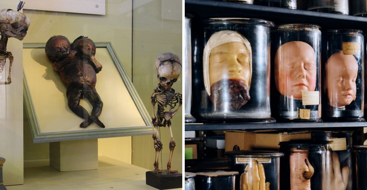
4. Conjoined Twins Preserved In Formaldehyde
Among the museum’s most emotionally powerful displays are several specimens of conjoined twins preserved in glass jars filled with formaldehyde. These infants, who shared body parts or organs, represent rare developmental conditions that occur approximately once in every 50,000 to 200,000 births. Medical professionals use these specimens to understand the complex embryological processes that sometimes go awry.
Each jar contains twins connected at different points, from shared livers to fused skulls, demonstrating the various ways this condition manifests. The preservation allows doctors to study internal organ placement and blood vessel connections that would be impossible to examine otherwise. Surgeons preparing for modern separation procedures have studied similar historical specimens to plan their approaches.
Many visitors experience conflicting emotions when viewing these displays, feeling both scientific curiosity and deep sadness for lives cut tragically short. The museum treats these specimens with dignity, providing educational context about the medical conditions rather than sensationalizing them. Parents often use this opportunity to discuss differences, empathy, and medical progress with their children in age-appropriate ways.
5. Slices Of Einstein’s Brain Tissue
Few people know that pieces of Albert Einstein’s actual brain reside in Philadelphia at the Mütter Museum. After the famous physicist died in 1955, pathologist Thomas Harvey removed his brain during autopsy, hoping to discover what made Einstein’s mind so extraordinary. Harvey sliced the brain into 240 pieces, preserving thin sections on microscope slides for scientific study.
The museum displays 46 of these slides, allowing visitors to peer at the cellular structure of genius itself. Researchers who studied Einstein’s brain found some unusual features, including extra folds in certain areas and a different ratio of neurons to support cells. However, scientists debate whether these differences truly explain his intellectual abilities or simply represent normal human variation.
Standing before these slides feels almost surreal, knowing you’re looking at tissue that once generated theories revolutionizing our understanding of space, time, and energy. Some critics questioned the ethics of removing Einstein’s brain without proper family consent. Nevertheless, these specimens continue sparking discussions about neuroscience, genius, and the physical basis of human thought and creativity.
6. The Mega Colon Measuring Five Feet
Prepare yourself for one of the museum’s most shocking displays featuring a human colon that grew to an absolutely enormous size. This specimen, removed from a patient suffering from Hirschsprung’s disease, measures approximately five feet long and weighs around 40 pounds when preserved. The condition occurs when nerve cells are missing from parts of the intestine, preventing normal muscle contractions that move waste through the digestive system.
As waste accumulated over years, the colon stretched to accommodate the blockage, eventually growing to grotesque proportions. The patient reportedly suffered terrible discomfort and health complications before doctors finally removed the diseased organ. Medical students study this specimen to understand severe gastrointestinal conditions and the importance of early diagnosis.
Visitors often gasp when they first see the megacolon because its size seems impossible for a human body part. The display includes explanatory text about the disease and modern treatment options, which typically involve surgery in infancy. This specimen powerfully illustrates how untreated medical conditions can drastically alter normal anatomy, making it both disturbing and educational for audiences of all ages.
7. Drawers Full Of Swallowed Objects
Opening certain museum drawers reveals a bizarre treasure trove of items that people accidentally or intentionally swallowed over the decades. Buttons, coins, safety pins, toy parts, and even small tools fill these collections, each object surgically removed from someone’s digestive tract. Some patients swallowed things by accident, while others suffered from pica, a condition causing people to eat non-food items.
Doctors in the 1800s and early 1900s lacked modern imaging technology, so they often discovered these objects only during surgery or autopsy. The collection shows how common this problem was before better safety standards for children’s toys and increased awareness of mental health conditions. Medical professionals today still occasionally remove strange objects from patients, though advanced endoscopic techniques make the procedures less invasive.
Kids especially love this exhibit because the objects look like random junk from a toy box or junk drawer. Parents use it as a teaching moment about choking hazards and why small objects don’t belong in mouths. The sheer variety of swallowed items makes you wonder about the stories behind each one and what circumstances led to such unusual accidents.
8. The Mummified Hand Of A Prisoner
Resting in a small case lies the mummified hand of a person believed to have been imprisoned or enslaved, still bearing marks that suggest restraints once bound the wrist. The hand dried out naturally through environmental conditions, creating a leathery, darkened appearance that preserved its shape and details. Fingernails, skin texture, and even fingerprint ridges remain visible despite the passage of time.
Historians and anthropologists study such specimens to learn about living conditions, labor practices, and treatment of marginalized people in past centuries. The restraint marks tell a silent story of suffering and loss of freedom that resonates powerfully with modern visitors. Museum curators present this specimen respectfully, acknowledging the human tragedy it represents rather than treating it as mere curiosity.
Many visitors pause here quietly, reflecting on historical injustices and the importance of human rights. Teachers bring students to discuss slavery, prison reform, and social justice using tangible historical evidence. The mummified hand serves as a sobering reminder that behind every medical specimen was a real person with experiences, emotions, and dignity that deserve recognition even centuries after death.
9. Facial Casts Of Famous Medical Cases
Before photography became widespread, doctors created plaster casts of patients’ faces to document unusual medical conditions, injuries, and diseases. The Mütter Museum houses an extensive collection of these haunting facial casts, including death masks and casts taken from living patients. Some show the devastating effects of untreated syphilis, while others display birth defects, tumors, or accident injuries.
Each cast captures incredible detail, from wrinkles and pores to the exact shape of lesions and deformities. Medical students in the 1800s studied these casts when they couldn’t observe patients directly, making them important teaching tools before modern medical imaging existed. The expressions frozen in plaster sometimes appear peaceful, while others seem to reflect pain or distress.
Walking past rows of disembodied faces creates an eerie atmosphere that makes visitors acutely aware of human vulnerability to disease and injury. Modern medicine can now treat or prevent many conditions shown in these casts, demonstrating how far healthcare has advanced. The collection also raises questions about patient consent and dignity, since many subjects likely had no choice about being documented for medical education purposes.
10. The Skeleton Of Conjoined Twins
Perhaps the museum’s most famous skeleton belongs to Chang and Eng Bunker, the original Siamese twins who lived from 1811 to 1874. Born in Siam (now Thailand), these brothers were connected at the chest by a band of cartilage and shared a fused liver. They traveled the world performing in exhibitions, eventually settling in North Carolina where they married sisters, fathered 21 children between them, and lived relatively normal lives.
After their deaths within hours of each other, doctors removed their connected liver and skeleton for medical study. The skeleton display shows exactly how their bodies were joined and how they managed to move and function together. Modern surgeons believe they could have been safely separated with today’s technology, but in their era, such surgery would have been fatal.
Their story challenges visitors to think about identity, brotherhood, and what it means to never be physically alone. The Bunker brothers succeeded in business, raised families, and lived to age 63, proving that physical differences don’t define a person’s potential. This exhibit remains deeply moving because it represents real people who turned their unique condition into a life of achievement and family connection.
Dear Reader: This page may contain affiliate links which may earn a commission if you click through and make a purchase. Our independent journalism is not influenced by any advertiser or commercial initiative unless it is clearly marked as sponsored content. As travel products change, please be sure to reconfirm all details and stay up to date with current events to ensure a safe and successful trip.